High-quality imaging systems are essential in today’s molecular biology, proteomics, and biomedical research. Whether you are quantifying proteins on Western blots, capturing faint chemiluminescent signals, or performing high-resolution tissue studies, a reliable imaging system ensures every experiment produces reproducible, publication-ready data.
Azure Biosystems offers a comprehensive range of imaging solutions—distributed and supported in South Africa by Apex Scientific—to meet the diverse needs of modern laboratories. This guide explores three core technologies: the compact chemiSOLO chemiluminescent imager, the versatile Western Blot Imaging Systems, and the multi-mode Sapphire FL/+ biomolecular imager. Together, these platforms cover applications from routine gel documentation to multiplex tissue analysis and in-vivo imaging.
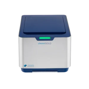
1. Chemiluminescent Imaging with the Azure chemiSOLO
Chemiluminescent Western blots remain a cornerstone of protein research. They allow scientists to detect extremely low-abundance proteins with clarity and precision. The Azure chemiSOLO brings this capability to any laboratory in a compact, user-friendly package.
Key Features
- Compact design without compromise – At only 29.2 × 22.2 × 43.2 cm, the chemiSOLO fits easily on crowded benches while maintaining high-performance imaging.
- High-resolution camera – A six-megapixel, back-illuminated CMOS sensor with Peltier cooling delivers low noise and exceptional sensitivity. A dynamic range greater than 2.1 optical density captures faint bands and intense signals in the same image.
- Browser-based control – Operate the instrument from any computer, tablet, or smartphone using a standard web browser. This simplifies setup and allows multiple researchers to access the system.
- Even illumination – Integrated transillumination LEDs provide uniform light for visible gel imaging and accurate background subtraction.
Applications
The chemiSOLO supports both routine and advanced workflows:
- Chemiluminescent Western blots with high signal-to-noise ratios.
- Visible protein gels stained with Coomassie or silver dyes.
- Quantitative densitometry for precise protein quantification when paired with the optional Densitometry Package (CS1004) and AzureSpot Pro software.
This combination of sensitivity, dynamic range, and ease of use allows South African laboratories to obtain reliable data quickly, whether documenting routine gels or publishing high-impact research.
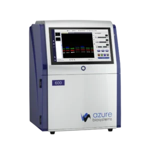
2. Western Blot Imaging Systems: End-to-End Workflow
Western blotting is a multi-step process in which every stage—electrophoresis, transfer, blocking, antibody incubation, and imaging—affects final data quality. Apex Scientific supplies Azure Western Blot Imaging Systems that integrate smoothly with consumables and accessories for consistent, reproducible results.
Step-by-Step Process
1. Gel electrophoresis – Proteins are first separated by size using SDS-PAGE. The Azure Aqua Quad Mini-Cell accommodates one to four gels (10 cm × 8 cm) and includes casting stands and frames. Researchers can run multiple samples simultaneously.
2. Membrane transfer – Separated proteins move to PVDF or nitrocellulose membranes using the Azure Aqua Transfer Cell. Color-coded components simplify setup, and integrated cooling units maintain optimal temperature. Selecting the correct membrane—PVDF for reprobing or nitrocellulose for high protein-binding affinity—ensures downstream detection accuracy.
3. Blocking non-specific binding – Blocking buffers such as Protein-Free Blot Blocking Buffer, Chemi Blot Blocking Buffer, or Fluorescent Blot Blocking Buffer reduce background noise and stabilize fluorescent signals. As a result, image clarity improves for both chemiluminescent and fluorescent detection.
4. Primary and secondary antibody incubation – After blocking, membranes are incubated with primary antibodies specific to the target protein, followed by secondary antibodies carrying detectable labels. Options include fluorescent secondary antibodies for visible or near-infrared fluorescence imaging and HRP-conjugated antibodies for chemiluminescent detection.
Azure’s Spectra Fluorescent Western Blotting Kits and chemiluminescent substrates—Radiance ECL, Radiance Plus, and Radiance Q—provide picogram- to femtogram-level sensitivity for rare protein targets.
5. Imaging and analysis – High-quality imaging is the final critical step. Azure Western Blot Imagers capture chemiluminescent, fluorescent, and visible gel signals with precision. Paired with AzureSpot Pro software, users can perform background subtraction, band detection, and total protein normalization using stains such as AzureRed or TotalStain Q. Automated analysis reduces variability and supports rigorous quantitative studies.
Choosing an system
When selecting a Western Blot Imaging System, consider:
- Detection modality—chemiluminescence, visible fluorescence, NIR fluorescence, or a combination.
- Sensitivity—especially important for low-abundance targets.
- Sample throughput—how many gels or membranes can be imaged simultaneously.
- Software capabilities—intuitive interfaces and robust quantitative tools.
By integrating compatible reagents, power supplies, and imaging systems, Apex Scientific ensures South African labs achieve reproducibility and efficiency from start to finish.
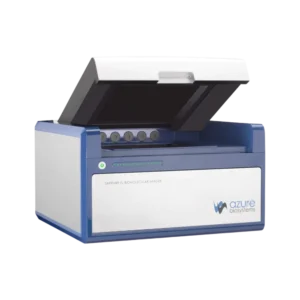
3. Sapphire FL/+ Biomolecular Imager: Multi-Mode, Ultra-High Resolution
For laboratories requiring the broadest range of applications, the Azure Sapphire FL/+ represents the pinnacle of imaging flexibility and sensitivity. This benchtop instrument combines point-scanning laser optics with optional sCMOS chemiluminescence detection, making it a true all-in-one platform.
Advanced Technology and Modular Design
- Point-scanning laser optics concentrate excitation light to a single point, producing exceptional signal-to-noise ratios and spatial resolution from 5 to 1 000 µm.
- Wide wavelength coverage from 375 to 900 nm accommodates UV through near-infrared dyes, enabling multiplex detection of up to four fluorescent targets simultaneously.
- Interchangeable laser modules and emission filters can be swapped in minutes, allowing laboratories to adapt the system to evolving experimental needs.
- Extended Dynamic Range (EDR) mode captures both weak and strong signals in a single scan for accurate quantification.
- Optional sCMOS camera adds sensitive chemiluminescent imaging without compromising fluorescence performance.
Versatile Applications
The Sapphire FL/+ supports fluorescence, chemiluminescence, and phosphor imaging on a single platform. It enables:
- Multiplex fluorescent Western blots without the need to strip and reprobe membranes.
- High-resolution gel imaging with uniform illumination for precise densitometry.
- Slide and tissue scanning for microarrays or tissue sections at up to 5 µm resolution.
- Phosphor imaging of radio-labelled samples with linear detection over more than five orders of magnitude.
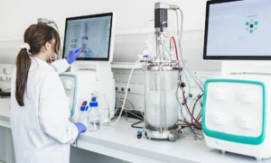
- In-vivo imaging of small animals, aided by five built-in anesthesia ports and a 4 cm imaging window for longitudinal studies.
Optional plate and slide holders enable high-throughput screening, making the Sapphire FL/+ suitable for research programs ranging from basic protein biology to preclinical drug development.
4. Bringing It All Together
Azure Biosystems imaging systems offer a scalable path from focused protein studies to complex, multiplexed imaging:
- The chemiSOLO provides cost-effective, high-sensitivity chemiluminescent imaging in a footprint small enough for any bench.
- Western Blot Imaging Systems deliver an integrated workflow for reproducible protein detection using chemiluminescence or fluorescence.
- The Sapphire FL/+ unites laser-based fluorescence, chemiluminescence, phosphor imaging, and in-vivo capability in a single high-resolution platform.
Each system is designed with researcher productivity in mind. Fast setup, intuitive operation, and powerful analysis software maintain the quantitative accuracy required for publication and regulatory compliance.
Conclusion
In modern life-science research, data quality depends on imaging systems that combine sensitivity, reproducibility, and flexibility. Whether you are documenting a single gel, analyzing multiple fluorescent targets, or imaging live animals, Azure Biosystems has a solution to meet your needs. With the expertise and local support of Apex Scientific, South African laboratories can confidently invest in imaging technology that not only meets today’s requirements but also adapts to tomorrow’s scientific challenges. From compact chemiluminescent imagers to state-of-the-art multi-mode platforms, Azure and Apex Scientific deliver the precision and reliability essential for breakthroughs in molecular biology and beyond.