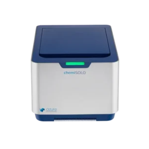
The western blot imaging is essential for proteomics, and Azure Biosystems’ imagers deliver high sensitivity with a mirror-free design. The CCD sensor sits directly above the sample to maximize light capture, boosting NIR fluorescence and chemiluminescent signal detection. In practice, this means researchers observe stronger signals and a wider dynamic range from each blot. Moreover, Apex Scientific provides these advanced laboratory imaging systems in South Africa, ensuring local labs have the latest equipment and support.
Mirrorless Direct Detection
Azure’s imagers use a mirror-free design that positions the camera sensor close to the sample. This direct detection approach greatly increases light collection efficiency, boosting sensitivity for even faint Western blot signals. As a result, more of the sample’s emitted light is captured compared to traditional systems with mirrors, improving data quality and quantification. Furthermore, placing the CCD sensor near the blot aligns with the inverse-square law (shown above), which enhances light capture. This design yields more reliable results across all Western blot imaging applications.
Enhanced Fluorescent Channel for Quantification
For precise quantification, Azure imagers offer an optional green fluorescence channel via the Q Module. The Azure Q upgrade adds a visible fluorescence path optimized for total protein stains (such as TotalStainQ), enabling accurate total protein normalization (TPN) of blots. In particular, using this TPN approach addresses the limitations of traditional housekeeping proteins at high loads, because the total-protein signal stays linear. Therefore, adding this visible fluorescence channel improves normalization accuracy and yields more dependable quantitative results.
Quantitative Chemiluminescent Imaging
Azure’s chemiluminescent detection rivals X-ray film sensitivity while enabling precise digital quantification. The system automatically detects and avoids CCD saturation (shown above) to ensure linearity across exposures, so quantitation remains accurate even for strong signals. As a result, researchers obtain publication-quality chemiluminescent images without a darkroom. Additionally, the system offers flexible controls. For example, users can:
- Change the shelf height to adjust the sample-to-lens distance for optimal focus.
- Choose preview, automatic, or manual capture modes as needed.
- Zoom on a region-of-interest (ROI) to minimize background noise and focus on key bands.
Overall, these features give complete control over exposure and enhance chemiluminescent workflow flexibility.
Multi-Channel Visible Fluorescence Imaging
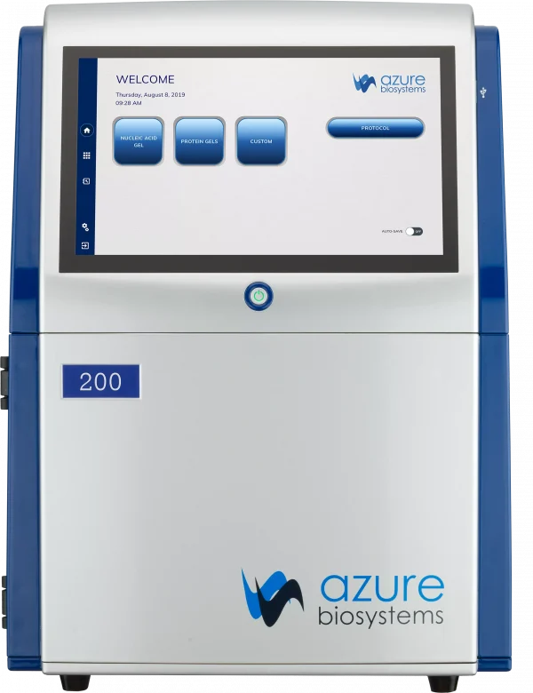
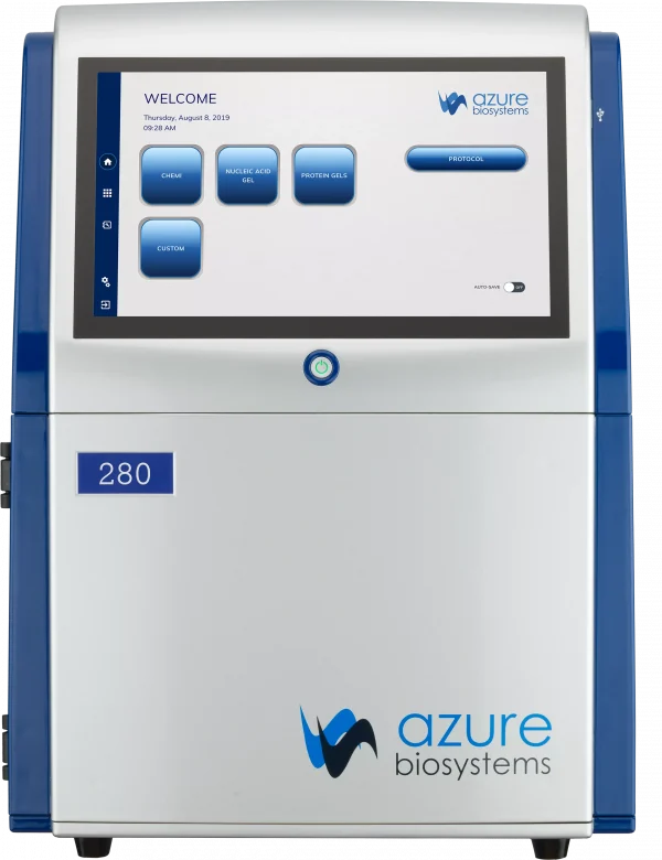
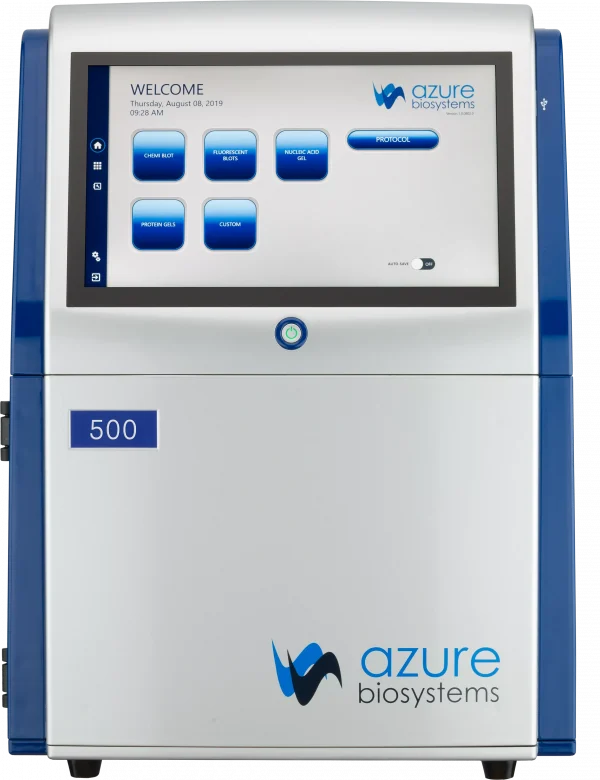
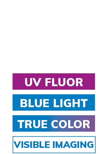
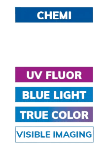
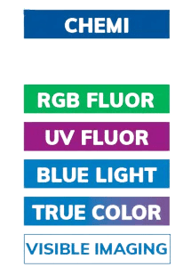
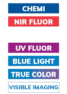
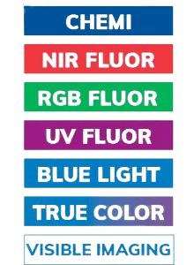
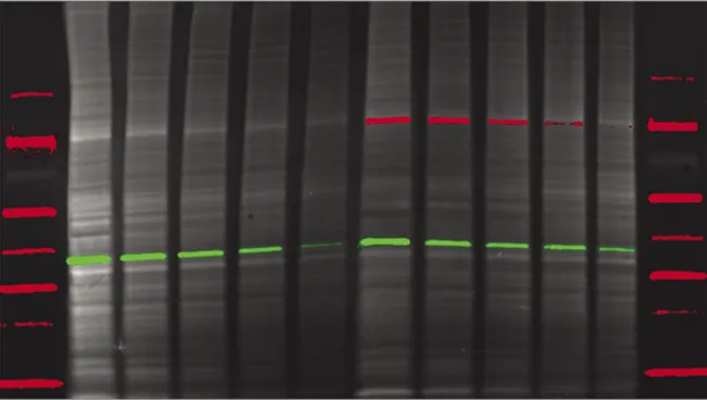
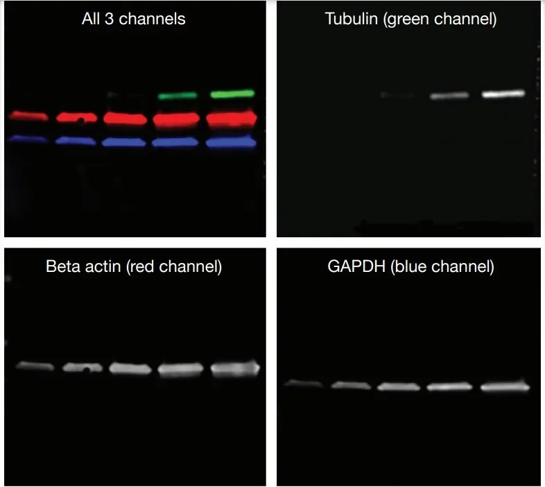
Azure imagers support full-color multiplex fluorescence from visible to NIR wavelengths. The built-in RGB LED illuminator and filters enable up to four fluorescent channels, so multiple targets can be imaged simultaneously. For example, the Azure 600 captures distinct red, green, and blue fluorescence channels in one blot (as shown above), allowing multiplex Western blots without changing setups. Users can choose an Azure 400 (visible-only), Azure 500 (NIR-only), or Azure 600 (both visible and NIR), each model expanding multiplex options within the same compact system.
Versatile Imaging Applications
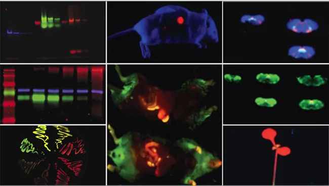
Azure imaging systems extend beyond blots to cover diverse applications. In addition to standard membranes and gel imaging, they handle membrane arrays (e.g. dot-blot grids) and visible gel stains with ease. For example, researchers routinely image gel electrophoresis results, cell culture plates, tissue sections, and even live small animals (mice, zebrafish) thanks to deep depth-of-field and high-resolution cameras (5.4–9.1 megapixels). Notably, entire three-dimensional samples can be scanned in situ, capturing volumetric fluorescence. This versatility means labs can rely on one instrument for many tasks, reducing the need for multiple specialized devices.
Broad Dye Compatibility
Azure imagers work with a wide range of fluorescent dyes and stains, ensuring compatibility with existing protocols. For example:
- Fluorescent dyes: Compatible with visible/NIR fluorophores like Alexa Fluor®, CyDyes, DyLight®, and Qdot® labels.
- Gel stains: Supports visible gel stains (e.g. Coomassie Blue, SYBR® Safe, Silver Stain, Ponceau) and fluorescent protein stains.
- Chemiluminescence: Captures ECL reagents and other luminescent substrates digitally.
In particular, near-infrared dyes (such as IRDye® 800CW) are detected with high sensitivity, and visible fluorescence markers (like Alexa Fluor 488 or SYPRO® Ruby) are easily imaged. The system’s multi-modal excitation (including UV, visible, and NIR LEDs) means a single instrument can handle most common stains. This broad compatibility ensures that laboratory protocols—whether chemiluminescent or fluorescent—work seamlessly with the Azure system.
For more detailed guidance and technical insights, visit the Apex Scientific Insights page.