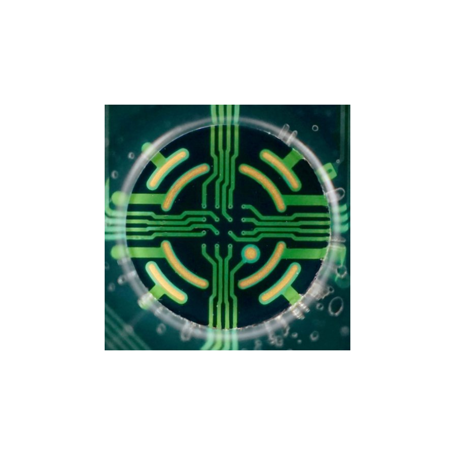
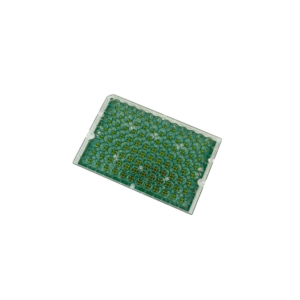
The BioCircuit microelectrode array (MEA) plates provide high-quality results together with industry-leading throughput at the lowest cost per well. The BioCircuit multiwell MEA plate is available in 24-, 48-, and 96-well formats. Featuring low-noise recording electrodes, dedicated stimulation electrodes (24- and 48-well versions only), and on-plate spotting guides to simplify plate preparation.
Key Features
High quality results with industry-leading throughput.
- Accurate cell spotting every time: BioCircuit MEA plates have on-plate spotting guides in the bottom of each well that confine the cell droplet over the recording electrodes.
- Record high quality data: BioCircuit MEA plates provide high-quality results together with industry-leading throughput at the lowest cost per well.
- Environmental control lids: The evaporation-reducing lids and built-in humidity chambers help controls your cells environment.
- Stimulate your cells: Each electrode is capable of both recording or stimulating your cells. In 24- and 48-well plates, each well contains a dedicated stimulation electrode for superior stimulation capacity and reliable capture.
| BioCircuit MEA
The BioCircuit MEA plates deliver high-quality results together with industry leading throughput at the lowest cost per well |
||||||||||
| Plate | Cat No. | Wells | Electrode/ well |
Electrode layout* |
Bottom | Walls | Maestro Edge |
Maestro Pro |
Maestro Z/ZHT |
Maestro Original |
|---|---|---|---|---|---|---|---|---|---|---|
| BioCircuit MEA 24 | M384-BIO-24 | 24 | 16 Gold |  |
Opaque | Clear | ● | ● | ||
| BioCircuit MEA 48 | M768-BIO-48 | 48 | 16 Gold |  |
Opaque | Clear | ● | ● | ||
| BioCircuit MEA 96 | M768-BIO-96 | 96 | 8 Gold |  |
Opaque | Clear | ● | ● | ||
*Schematic of well illustrating recording electrodes (blue), grounds (orange), and where present, a large dedicated stimulation (blue).
Overview
Superior droplet placement
BioCircuit MEA plates have on-plate spotting guides in the bottom of each well that confine the cell droplet over the recording electrodes. This enables more precise cell plating with less effort. Simply position the pipette between the AccuSpot features and release the droplet to ensure a perfectly centered, rounded droplet in every well.Plating cells in a small droplet centered over the electrode array will conserve cells and ensure robust electrical activity near the electrodes.
High quality MEA data
All excitable cell types perform well on the BioCircuit MEA plates, showing excellent coverage across all wells and the high signal-to-noise ratio. Track the development of electrical activity in stem cell-derived neurons or cardiomyocytes, study disease models, or examine the effects of compound treatment on excitable cells – the possibilities are endless with BioCircuit MEA plates.
Enhance your assay with LEAP
To complement high-quality field potential signals recorded using BioCircuit MEA plates, Axion’s patent-pending Local Extracellular Action Potential (LEAP) assay allows you to record extracellular action potential waveforms from cardiomyocytes. LEAP signals enable quantification of action potential morphology and characterization of complex repolarization irregularities such as early after depolarizations (EADs).
Related Applications
- Cardiac Classification – Predict the cardiomyocyte subtype using the action potential waveform in the LEAP assay
- Cardiac Development – Track cardiac differentiation and maturation over weeks and determine when cardiomyocytes start to beat and form a syncytium
- Cardiomyocyte Pacing – Control cardiomyocyte beat rate with optical or electrical stimulation to increase the physiological relevance of the assay
- Cardiotoxicity and Safety – Evaluate the proarrhythmic risk of compounds in acute and chronic exposure experiments in CiPA-style studies
- Neural Characterization and Development – Monitor neural development and quantify network dynamics to identify unique properties of your culture
- Neural Co-culture and Glial Interaction – A wide variety of neurons and other cells exist and interact together and the composition of a neural culture can alter development and disease progression.
- Neural Optical and Electrical Stimulation – Evoke neural activity with optical or electrical stimulation
- Neural Organoids – Measure neural activity from live neural organoids and characterize advanced network behavior
- Neurological Disease – Explore the role your gene of interest has on neural network activity in patient-derived iPSCs or animal models
- Neuromuscular Junction – Record evoked skeletal myotube activity in functional iPSC-derived neuromuscular junctions
- Neurotoxicology and Safety – Characterize the neuroactive properties of compounds in acute and chronic exposure experiments
- Pain – Measure modulations of DRG neuron activity in response to chemical or temperature stimuli
- Retina – Record light-evoked electrical activity from primary retina slices and iPSC-derived retinal organoids
- Zebrafish – Track neural activity, from zebrafish brain and spinal cord, label free


